مجموعة بيانات أورام الدماغ
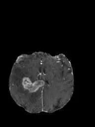
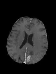
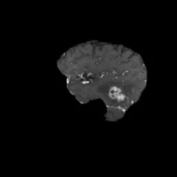
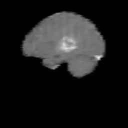

مجموعة بيانات أورام الدماغ من Ultralytics هي مجموعة بيانات لاكتشاف الكائنات تضم 1,115 صورة للتصوير بالرنين المغناطيسي للدماغ (994 للتدريب و121 للتحقق)، مع 1,180 صندوق إحاطة ضمن فئة واحدة، هي tumor. تحتوي كل صورة على صندوق إلى 3 صناديق للأورام. يضم قسم التحقق صور 25 مريضًا جرى تحديد هويتهم، ولا يظهر أي مريض معروف في كلا القسمين. ويبلغ حجم المجموعة 4.06 MB، وهي مجموعة مدمجة لتعلّم نماذج الرؤية الحاسوبية وإنشاء نماذج أولية لـتطبيقات الرعاية الصحية، وليست موردًا تشخيصيًا معتمدًا سريريًا.
شاهد: اكتشاف أورام الدماغ باستخدام منصة Ultralytics وUltralytics YOLO26 | اكتشاف الكائنات 🚀
بنية مجموعة البيانات#
تضم مجموعة بيانات أورام الدماغ من Ultralytics عدد 1,115 صورة للتصوير بالرنين المغناطيسي للدماغ و1,180 صندوق إحاطة في قسمين يحددهما التكوين brain-tumor.yaml:
| المجموعة الفرعية | الصور | صناديق الإحاطة | المرضى |
|---|---|---|---|
| التدريب | 994 | 1,057 | 95 مريضًا بمعرّف، إضافة إلى 541 صورة بلا معرّف للمريض |
| التحقق | 121 | 123 | 25 |
| الإجمالي | 1,115 | 1,180 |
تحتوي كل صورة على صندوق إلى 3 صناديق إحاطة من الفئة الوحيدة tumor، وتتراوح أبعاد الصور من 192x256 إلى 512x512 بكسل. ولا يحدد التكوين قسم اختبار منفصلًا.
أُنشئ قسم التحقق بحسب المرضى. تبدأ بعض أسماء الملفات بمعرّف مريض من خمسة أرقام؛ فعلى سبيل المثال، ينتمي 00097_77.jpg إلى المريض 00097. تشكّل الصور الحاملة لمعرّفات 25 مريضًا من هؤلاء قسم التحقق، بينما توجد صور المرضى الـ95 الآخرين المعروفين في قسم التدريب، لذا لا يظهر أي مريض معروف في كلا القسمين. أما الصور الـ541 المتبقية، فلا تحمل أسماء ملفاتها معرّفًا للمريض، وتوجد جميعها في قسم التدريب.
تُنزّل مجموعة البيانات تلقائيًا (4.06 MB) من أصول Ultralytics على GitHub عند التدريب للمرة الأولى، لذا لا حاجة إلى تنزيلها أو إعدادها يدويًا.
تغيّرت مجموعة البيانات إلى فئة واحدة tumor في نوفمبر 2026. إذا كان مجلد brain-tumor/images/train المحلي لديك يحتوي على 893 صورة بدلًا من 994، فاحذف مجلد brain-tumor من دليل مجموعات البيانات (راجع datasets_dir في yolo settings) وحدّث الحزمة باستخدام pip install -U ultralytics. بعد ذلك، تُنزّل النسخة الحالية عند تشغيل التدريب التالي.
استكشف أورام الدماغ على منصة Ultralytics لتصفح الصور مع طبقات التعليقات التوضيحية، وعرض توزيع الفئات والخرائط الحرارية لمربعات الإحاطة في علامة التبويب Charts، واستنساخ المجموعة لتدريب نموذجك الخاص سحابيًا.
التطبيقات#
تحدد نماذج الكشف عن أورام الدماغ مواقع الأورام في صور التصوير بالرنين المغناطيسي للدماغ. وبفضل 1,115 صورة وحجم تنزيل يبلغ 4.06 MB، تناسب مجموعة بيانات أورام الدماغ من Ultralytics ما يلي:
- تعلّم سير العمل: تنفيذ دورة التدريب والتحقق والتنبؤ كاملةً على صور طبية باستخدام مجموعة بيانات صغيرة.
- إنشاء النماذج الأولية: بناء مسار لتحديد مواقع الأورام واختباره قبل الانتقال إلى مجموعة بيانات سريرية أكبر.
- التقييم على مرضى معروفين: قياس أداء الكشف على المرضى الـ25 في قسم التحقق. ولأن 541 صورة تدريب لا تحمل معرّفًا للمريض، فلا يمكن استبعاد احتمال تداخلها مع صور هؤلاء المرضى.
- البحث والتعليم: مقارنة نماذج الكشف ودراسة تحليل الصور الطبية والذكاء الاصطناعي في الأشعة.
النماذج المدرّبة على مجموعة البيانات هذه مخصصة للبحث والتعليم، ولم تُعتمد للتشخيص السريري.
ملف YAML لمجموعة البيانات#
يحدد التكوين brain-tumor.yaml مسار مجموعة البيانات ومجلدي صور التدريب والتحقق والفئة الوحيدة tumor وعنوان URL للتنزيل. وتستخدم التسميات تنسيق Ultralytics YOLO.
# Ultralytics 🚀 AGPL-3.0 License - https://ultralytics.com/license

# Brain-tumor dataset by Ultralytics
# Documentation: https://docs.ultralytics.com/datasets/detect/brain-tumor
# Example usage: yolo train data=brain-tumor.yaml
# parent
# ├── ultralytics
# └── datasets
#     └── brain-tumor ← downloads here (4.06 MB)

# Train/val/test sets as 1) dir: path/to/imgs, 2) file: path/to/imgs.txt, or 3) list: [path/to/imgs1, path/to/imgs2, ..]
path: brain-tumor # dataset root dir
train: images/train # train images (relative to 'path') 994 images
val: images/val # val images (relative to 'path') 121 images

# Classes
names:
  0: tumor

# Download script/URL (optional)
download: https://github.com/ultralytics/assets/releases/download/v0.0.0/brain-tumor.zipالاستخدام#
لتدريب نموذج YOLO26n على مجموعة بيانات أورام الدماغ لمدة 100 حقبة وبحجم صورة 640، استخدم مقتطفات الشيفرة أدناه.
from ultralytics import YOLO
# تحميل نموذج
model = YOLO("yolo26n.pt") # تحميل نموذج مدرّب مسبقًا (موصى به للتدريب)
# تدريب النموذج
results = model.train(data="brain-tumor.yaml", epochs=100, imgsz=640)from ultralytics import YOLO
# تحميل نموذج
model = YOLO("path/to/best.pt") # حمّل نموذج أورام الدماغ محسّن الضبط
# الاستدلال باستخدام النموذج
results = model.predict("https://ultralytics.com/assets/brain-tumor-sample.jpg")صور وتعليقات توضيحية نموذجية#
فيما يلي صور نموذجية من مجموعة بيانات أورام الدماغ من Ultralytics مع تسمياتها التوضيحية:
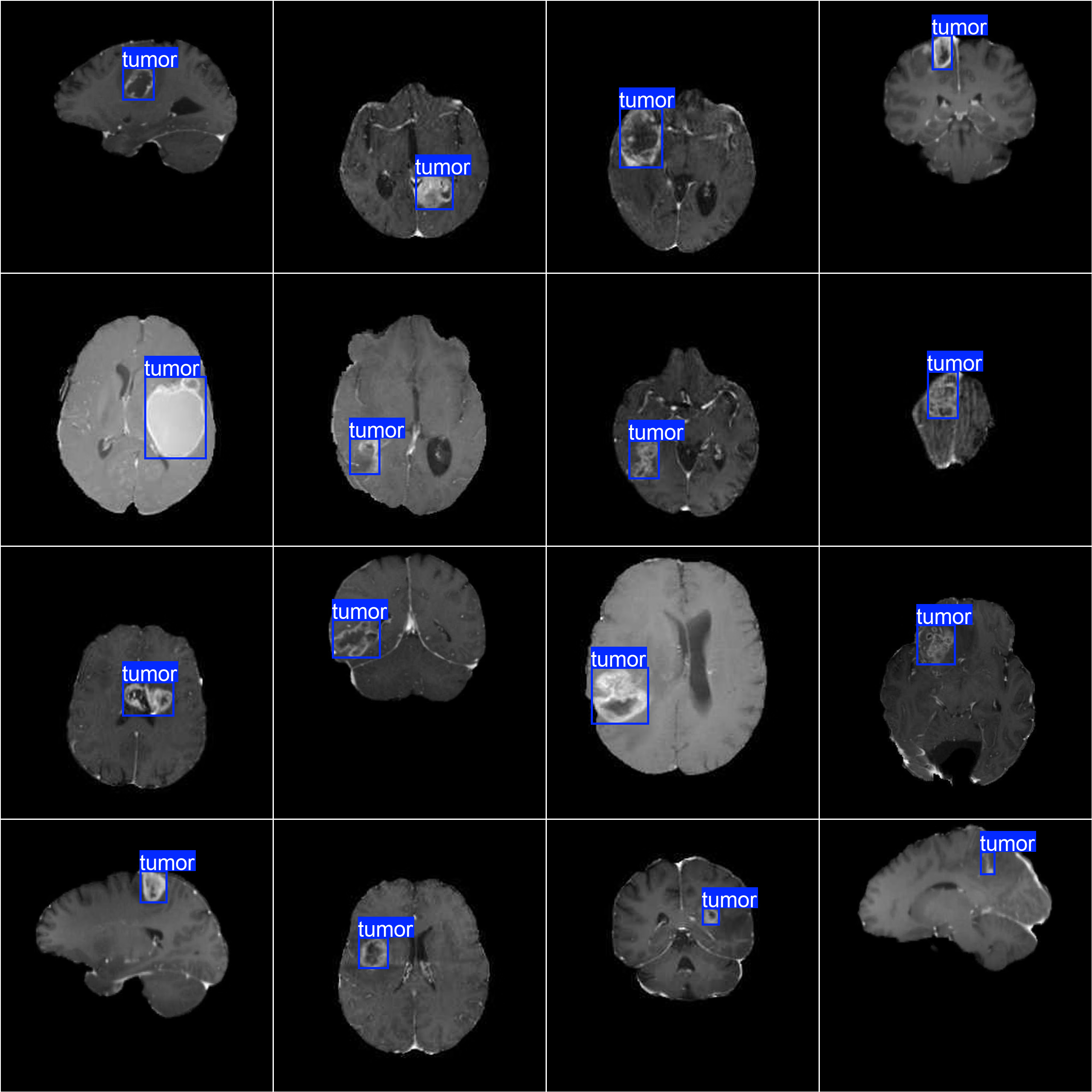
- الصور الموسومة: تعرض الشبكة 16 صورة تدريب، تحتوي كل منها على صندوق إحاطة
tumorحول الآفة. وتختلف الأورام في الحجم والشكل والموقع ضمن مجموعة البيانات، ما يساعد نموذج الكشف على التعميم.
الاستشهادات والشكر والتقدير#
مجموعة البيانات متاحة بموجب ترخيص AGPL-3.0.
إذا استخدمت مجموعة بيانات أورام الدماغ في عملك البحثي أو التطويري، فيُرجى الاستشهاد بها:
@dataset{Ultralytics_Brain_Tumor_Dataset_2023,
    author = {Ultralytics},
    title = {Brain Tumor Detection Dataset},
    year = {2023},
    publisher = {Ultralytics},
    url = {https://docs.ultralytics.com/datasets/detect/brain-tumor}
}
الأسئلة الشائعة#
تضم مجموعة بيانات أورام الدماغ من Ultralytics عدد 1,115 صورة للتصوير بالرنين المغناطيسي للدماغ وفئة واحدة، هي
tumor: 994 صورة تدريب مع 1,057 صندوق إحاطة، و121 صورة تحقق مع 123 صندوق إحاطة. تحتوي كل صورة على صندوق واحد إلى 3 صناديق، ولا يوجد قسم اختبار منفصل. راجع بنية مجموعة البيانات للاطلاع على تفاصيل التقسيم.تحتوي مجموعة بيانات أورام الدماغ من Ultralytics على صور للتصوير بالرنين المغناطيسي للدماغ، ولا تتضمن صورًا للتصوير المقطعي المحوسب. وتتراوح أبعاد الصور من 192x256 إلى 512x512 بكسل، وتحمل كل صورة من صندوق إلى 3 صناديق إحاطة
tumorوفق تنسيق Ultralytics YOLO.لا، لا تحتوي مجموعة بيانات أورام الدماغ من Ultralytics على صور خالية من الأورام ولا على فئة سلبية. وتضم الصور الـ1,115 كلها من صندوق إلى 3 صناديق إحاطة
tumor، لذا يتعلم النموذج المدرّب عليها موضع الورم، لا ما إذا كان الفحص سليمًا. والنماذج المدرّبة على هذه المجموعة مخصصة للبحث والتعليم، لا للتشخيص السريري.تُقسّم مجموعة بيانات أورام الدماغ من Ultralytics بحسب المرضى: تحمل صور التحقق الـ121 معرّفات 25 مريضًا، ولا يظهر أي مريض معروف في كلا القسمين. ويُحدَّد المرضى بواسطة البادئة ذات الأرقام الخمسة في اسم الملف، مثل
00097_77.jpg. تضم مجموعة التدريب المرضى الـ95 الآخرين المعروفين، إضافةً إلى 541 صورة لا تحمل أسماء ملفاتها معرّفًا للمريض.تُنزّل مجموعة بيانات أورام الدماغ من Ultralytics (4.06 MB) تلقائيًا من أصول Ultralytics على GitHub عند التدريب للمرة الأولى باستخدام
data="brain-tumor.yaml"، لذا لا حاجة إلى تنزيلها يدويًا. إذا كانت نسخة أقدم تضم 893 صورة تدريب موجودة على القرص، فاحذف مجلدbrain-tumorوحدّث حزمةultralyticsأولًا لتنزيل النسخة الحالية ذات الفئة الواحدة. وللاطلاع على مجموعة بيانات تصوير طبي صغيرة أخرى، راجع مجموعة بيانات الكشف عن الحبوب الطبية.لتدريب YOLO26 على مجموعة بيانات أورام الدماغ من Ultralytics، مرّر
data="brain-tumor.yaml"إلى أمر التدريب، وستُنزّل مجموعة البيانات تلقائيًا. تدرّب الأمثلة أدناه YOLO26n لمدة 100 حقبة بحجم صورة يبلغ 640:مثال على التدريبfrom ultralytics import YOLO # تحميل نموذج model = YOLO("yolo26n.pt") # تحميل نموذج مدرّب مسبقًا (موصى به للتدريب) # تدريب النموذج results = model.train(data="brain-tumor.yaml", epochs=100, imgsz=640)للاطلاع على قائمة مفصلة بالوسائط المتاحة، راجع صفحة التدريب.
لإجراء الاستدلال باستخدام نموذج YOLO26 مضبوط بدقة على مجموعة بيانات أورام الدماغ من Ultralytics، حمّل أوزان
best.ptواستدعِ predict على صورة للتصوير بالرنين المغناطيسي للدماغ. تحتوي كل نتيجة على صناديق الإحاطةtumorمع درجات الثقة:مثال على الاستدلالfrom ultralytics import YOLO # تحميل نموذج model = YOLO("path/to/best.pt") # حمّل نموذج أورام الدماغ محسّن الضبط # الاستدلال باستخدام النموذج results = model.predict("https://ultralytics.com/assets/brain-tumor-sample.jpg")أنشأت Ultralytics مجموعة بيانات أورام الدماغ، وهي متاحة بموجب ترخيص AGPL-3.0.


التعليقات11
اطرح سؤالًا، وشارك نتائجك، أو ساعد شخصًا آخر.
ZoltinZyc
how can i download this datasett??
1 Paula DerrengerUltralytics(تم التعديل)
Hey there! 😊 The easiest way is to train with
data=brain-tumor.yaml, which downloads the dataset automatically the first time. To download it manually, use:wget https://github.com/ultralytics/assets/releases/download/v0.0.0/brain-tumor.zip unzip brain-tumor.zip -d path/to/destinationReplace
path/to/destinationwith the directory where you'd like the dataset stored. Once you have the dataset, you're all set to start training or inferencing with YOLO models on brain tumor images!Should you need further guidance, feel free to ask. Happy coding! 🚀
G-qingqian
Thank you for your reply. I noticed that in the original dataset, the images in the train folder do have ID prefixes, but the images in the val folder are all prefixed with val1_, making it impossible to distinguish which patient the data belongs to. Therefore, I cannot redivide the dataset. Could you please provide data with complete identifiers? ---- Replied Message ---- FromGlenn @.>Date1/18/2026 @.>@.>, @.>SubjectRe: [ultralytics/ultralytics] zh/datasets/detect/brain-tumor/ (Discussion #9988) The packaged brain-tumor dataset split is provided at the image-folder level and doesn’t ship patient IDs/metadata, so we can’t guarantee a patient-disjoint (no same patient in both train/val) split; if you need patient-wise separation, you’ll need to rebuild the split from patient identifiers and point a custom YAML to *.txt image lists (supported by Ultralytics YOLO), e.g. see the provided config in the Brain Tumor dataset docs and the brain-tumor.yaml in the repo.
my-brain-tumor.yaml path: /data/brain-tumor train: splits/train.txt # one image path per line (ideally grouped by patient ID when you create it) val: splits/val.txt names: 0: negative 1: positive
— Reply to this email directly, view it on GitHub, or unsubscribe. You are receiving this because you commented.Message ID: @.***>
 Glenn JocherUltralytics
The packaged
brain-tumorzip only includes image-level labels with anonymized filenames (no patient IDs/metadata), so we can’t provide “complete identifiers” or guarantee a patient-disjoint re-split; for patient-wise splitting you’ll need to rebuild from a source that includes patient IDs (e.g., your DICOM/NIfTI originals) and then generatetrain.txt/val.txt, and if you just need stronger evaluation without IDs you can use our K-Fold Cross Validation guide—please keep follow-ups here in the GitHub discussion (we can’t provide support over email/DM).
Can your dataset ensure that the data are independently and identically distributed by patient? That is, there will be no data from the same patient in both the training set and the validation set.
- Glenn JocherUltralytics
The packaged
brain-tumordataset split is provided at the image-folder level and doesn’t ship patient IDs/metadata, so we can’t guarantee a patient-disjoint (no same patient in both train/val) split; if you need patient-wise separation, you’ll need to rebuild the split from patient identifiers and point a custom YAML to*.txtimage lists (supported by Ultralytics YOLO), e.g. see the provided config in the Brain Tumor dataset docs and the brain-tumor.yaml in the repo.# my-brain-tumor.yaml path: /data/brain-tumor train: splits/train.txt # one image path per line (ideally grouped by patient ID when you create it) val: splits/val.txt names: 0: negative 1: positive
pizzas17
How could I download this dataset on COCO format or how could I convert it? (Since I wish to have an EDA on it
xySun17
How can i train the yoloV8 using this dataset without loading the pre-trained weights?
- Paula DerrengerUltralytics(تم التعديل)
@xySun17 hello!
To train YOLO26 without loading the pre-trained weights, you can initialize the model from scratch by specifying the model architecture file (e.g.,
yolo26n.yaml) instead of the pre-trained weights file. Here’s how you can do it:Python
from ultralytics import YOLO

# Load a model architecture without pre-trained weights
model = YOLO("yolo26n.yaml")  # build a new model from YAML

# Train the model
results = model.train(data="brain-tumor.yaml", epochs=100, imgsz=640)
CLI
# Train from scratch using the model architecture file
yolo detect train data=brain-tumor.yaml model=yolo26n.yaml epochs=100 imgsz=640
This approach ensures that the model starts training with random weights rather than pre-trained ones. If you encounter any issues, please ensure you are using the latest version of the Ultralytics package and provide a minimum reproducible example if the problem persists. You can find more details on creating a reproducible example here.
Feel free to reach out if you have any further questions. Happy training! 🚀
1 Thank you!! However, there is another problem, that is, why it says the path of dataset is not found? I already downloaded the dataset files and changed the path in "brain-tumor.yaml" file.
The threw error was follows: "Dataset 'brain-tumor.yaml' images not found ⚠️, missing path '/valid/images' Note dataset download directory is '/home/ros/datasets'. You can update this in '/home/ros/.config/Ultralytics/settings.yaml'"
- Paula DerrengerUltralytics(تم التعديل)
Hello @xySun17,
Thank you for reaching out! It looks like the issue might be related to the dataset path configuration in your
brain-tumor.yamlfile. Here are a few steps to troubleshoot and resolve this:-
Verify Dataset Path: Ensure that the paths specified in your
brain-tumor.yamlfile correctly point to the locations of your dataset images. For example:path: /home/ros/datasets/brain-tumor train: images/train val: images/val -
Check Directory Structure: Confirm that the directory structure matches what is specified in the YAML file. The
images/trainandimages/valdirectories should exist within/home/ros/datasets/brain-tumor. -
Update Configuration: If the dataset is located in a different directory, update the
pathin the YAML file accordingly. For instance, if your dataset is in/home/ros/my-datasets/brain-tumor, update thepathto:path: /home/ros/my-datasets/brain-tumor -
Reproducible Example: If the issue persists, providing a minimum reproducible example can greatly help in diagnosing the problem. You can find guidance on creating a reproducible example here.
-
Latest Version: Ensure you are using the latest version of the Ultralytics package. You can update it using:
pip install --upgrade ultralytics
Here’s an example of how your
brain-tumor.yamlshould look:# Ultralytics 🚀 AGPL-3.0 License - https://ultralytics.com/license # Brain-tumor dataset by Ultralytics # Documentation: https://docs.ultralytics.com/datasets/detect/brain-tumor/ # Example usage: yolo train data=brain-tumor.yaml path: /home/ros/datasets/brain-tumor # dataset root dir train: images/train # train images (relative to 'path') val: images/val # val images (relative to 'path') # Classes names: 0: negative 1: positive # Download script/URL (optional) download: https://github.com/ultralytics/assets/releases/download/v0.0.0/brain-tumor.zipIf you follow these steps and still encounter issues, please share more details or any error messages you receive. This will help us assist you better.
Best regards and happy coding! 🚀
-